The importance of optimal preparation quality in Digital Cytology in the screening for Cervical Cancer.
Digital Cytology is more and more introduced in the screening for Cervical Cancer.
However, working with Digital Cytology (even for conventional screening method!) requires a high standard of quality of the microscopic slides produced.
Standardization, in the pre-processing, is key in the success of optimal results in the screening for Cervical Cancer, from sample taking to scanning the LBC-slides.
To all sample takers:
“An optimal sample collection is the most important part for having an accurate diagnosis”
An optimal PAP-smear consists of:
Cells from the transformation zone (see the importance in CASE N°12)
NO contamination by lubricants and ultrasonography gels, as these products have a huge impact on the quality and quantity of the processed slides and therefor on the diagnosis.
Only water-soluble carbomer-free gels lubricant sparingly applied to the posterior blade of the speculum can be used if necessary!
To all cytology lab-technicians:
“An optimally manufactured slide, will provide a more accurate and reliable diagnosis”
How to get an optimal manufactured slide:
Work standardized
Follow carefully the requirements of the Liquid Based Cytology manufacture
Follow exactly the staining protocol by using the required dyes, solutions and ethanol dilutions
Ensure that the slides are optimally dehydrated to prevent the cornflakes artifact.¹
Check if there is no residue of cover-slipping medium, dust or fingerprints on the cover-slipping or even air-bubbles under the cover-slipping.
Digital Cytology is not Science Fiction: garbage will not change into gold!
To all CytoTechnologists and Cyto-Pathologists:
CytoProcessor will aid you to make accurate and reliable diagnoses with well prepared, optimal stained and good cover-slipped LBC-slides.
Let’s do it together for the women behind the PAP-smear!
— Annette W. Hamminga (CTIAC), Digital Cytology Expert at Datexim
¹ Read the article: BRIEF REPORT - Causation of cornflake artifacts
CytoProcessor® is a CE marked IVD device, intended for use by healthcare professionals only. Please consult the user guide provided by the manufacturer, DATEXIM, for detailed conditions of use and recommendations for safe and effective operation.
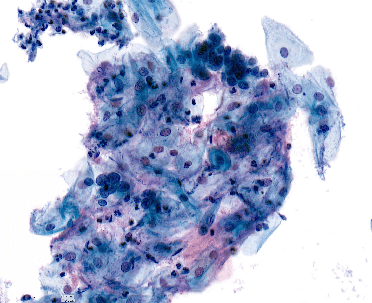
PICTURE 1
Optimal preparation and staining results
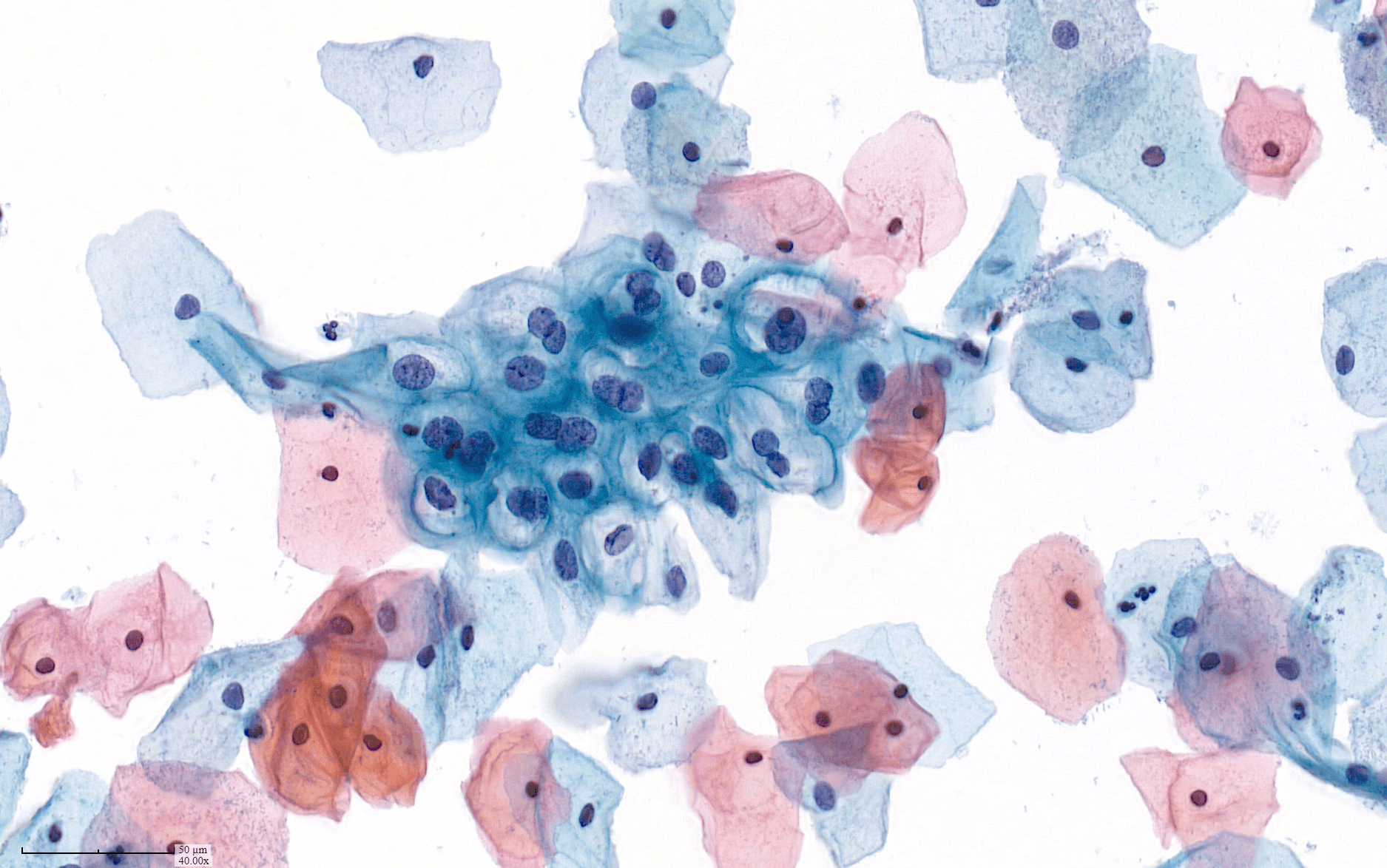
PICTURE 2
Ultrasonography gel
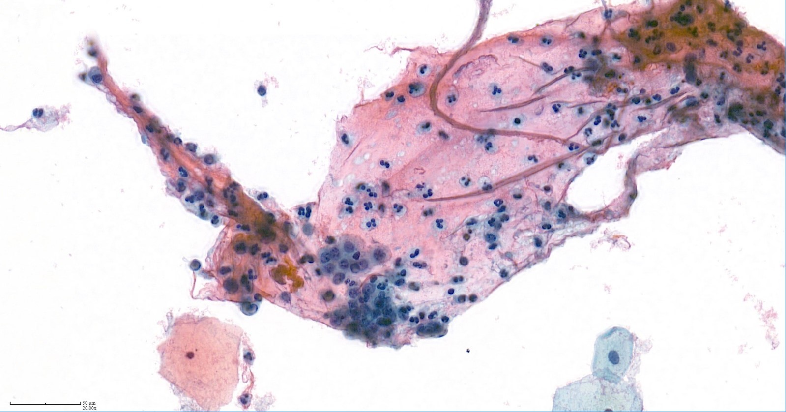
PICTURE 3
HSIL cells captured in gel and polymorphs

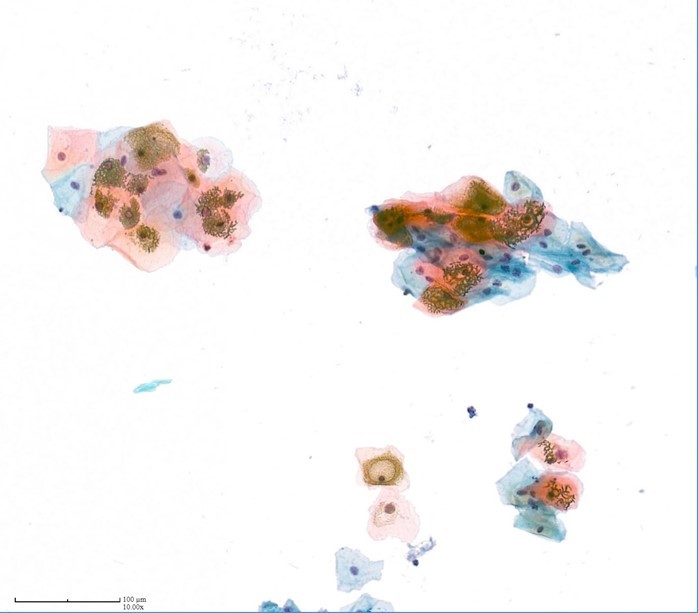
PICTURE 4
Cornflakes artifacts
Link to others articles :

